Examinations of the anterior segment of the eye
OCT of the cornea, sclera, and iridocorneal angle
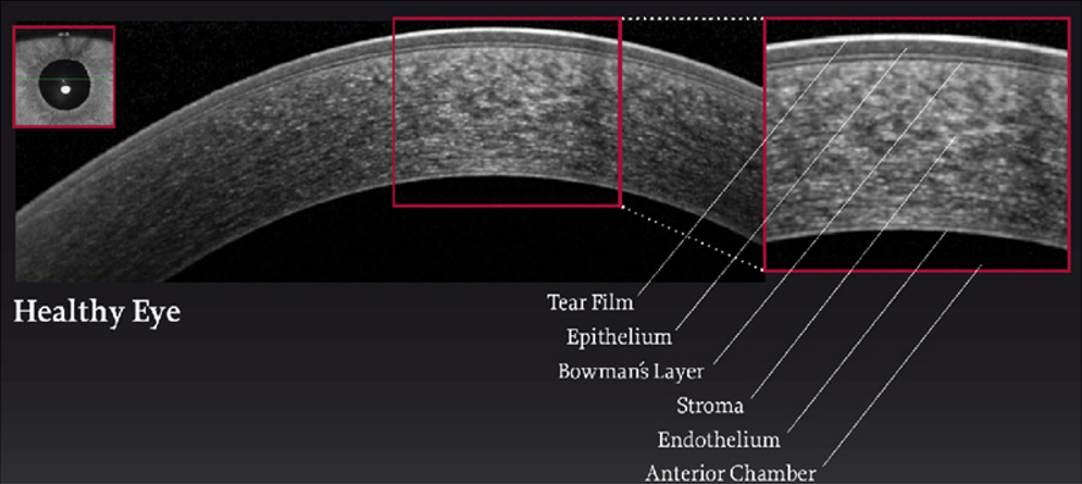
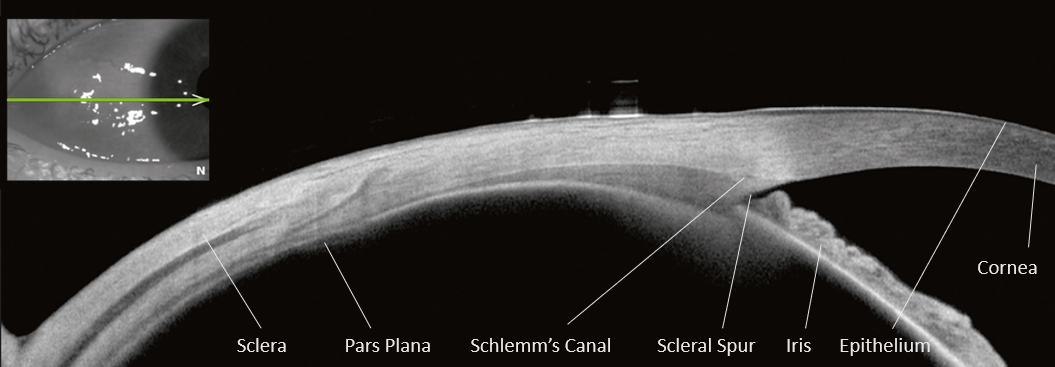
Optical coherence tomography (OCT) can be used to view the cornea, sclera, or corneal angle as a cross-section. A sensitive, safe laser scans the corresponding structure without touching it and displays it as a cross-section.
The OCT of the cornea does not require the administration of drugs, is painless, and takes only a few minutes for each eye.
Measurement of the anterior segment of the eye
An OCT adapted to a slit lamp can measure the corneal radius and the central corneal thickness. Likewise, the depth (= distance from the posterior corneal surface to the eye lens) and the volume of the entire anterior chamber can be determined.
A slit lamp is a microscope used by the examiner to view the eye at many times the magnification. The microscope has a light source that can be swiveled to both sides. This light produces a slit-shaped, focused light beam that shines through the transparent parts of the eye. The eyecare professional moves this light beam slowly across the eye at the appropriate magnification. Any changes, such as on the cornea or the lens, become visible.
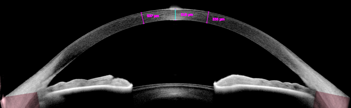
Pachymetry (thickness measurement) of the cornea
With corneal pachymetry, the examiner obtains a quick overview of the cornea’s thickness at any point. This allows the measurement of structural changes in the cornea. The thickness of the cornea is an important parameter for carrying out refractive surgery (e.g., LASIK), the surgery that can correct your vision.
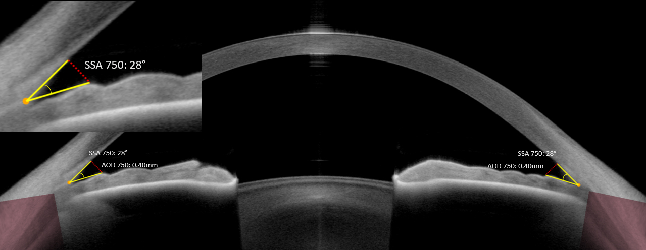
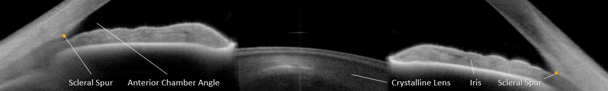
Digital gonioscopy
Digital gonioscopy is the examination of the corneal angle in the eye using an OCT instrument. Angles occur on both sides of the eye where the cornea and the iris come together. Extra fluid drains from the anterior chamber of the eye via these angles. If these drainage paths are blocked, this may result in increased eye pressure and thus in serious visual disorders. For this reason, it is important to observe, evaluate, and measure the angles.